Spatial Angle Imaging
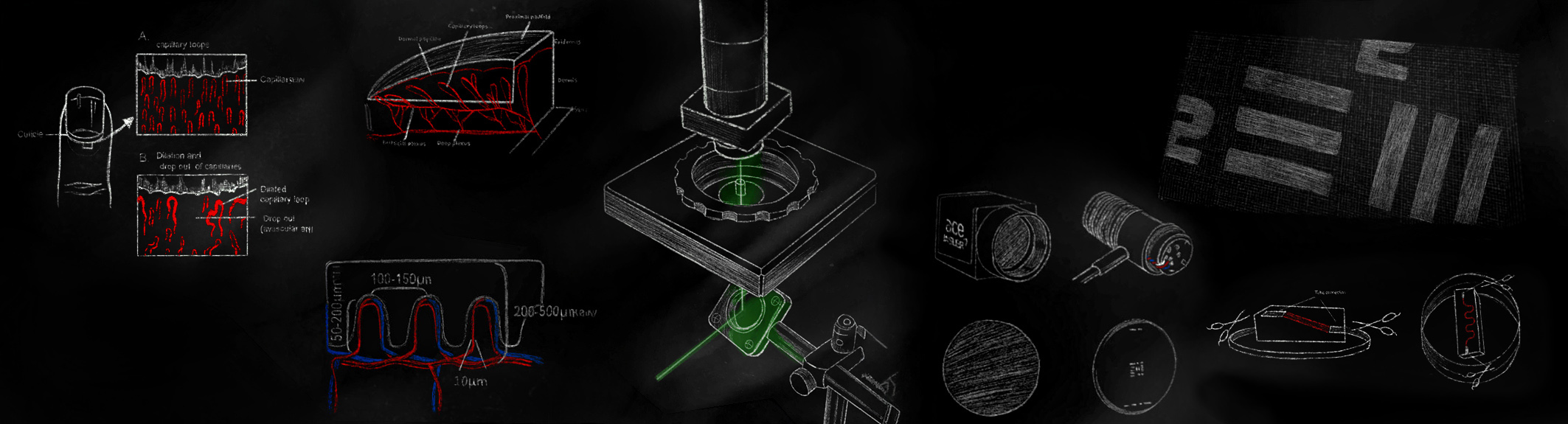
This project involves developing optical probes that enable deeper visual penetration into biological tissues, where light scattering is a significant limitation. The Spatial Angle Filtering (SAF) technology uses a specialized fibre-optic plate (FOP) to reject scattered photons from the tissue while preserving photons carrying subcutaneous visual information. This research aims to address the gap in capillary loop imaging (which is significant for detecting superficial cancers) by proposing an optimized imaging platform that enhances resolution, penetration depth, portability, and cost efficiency.
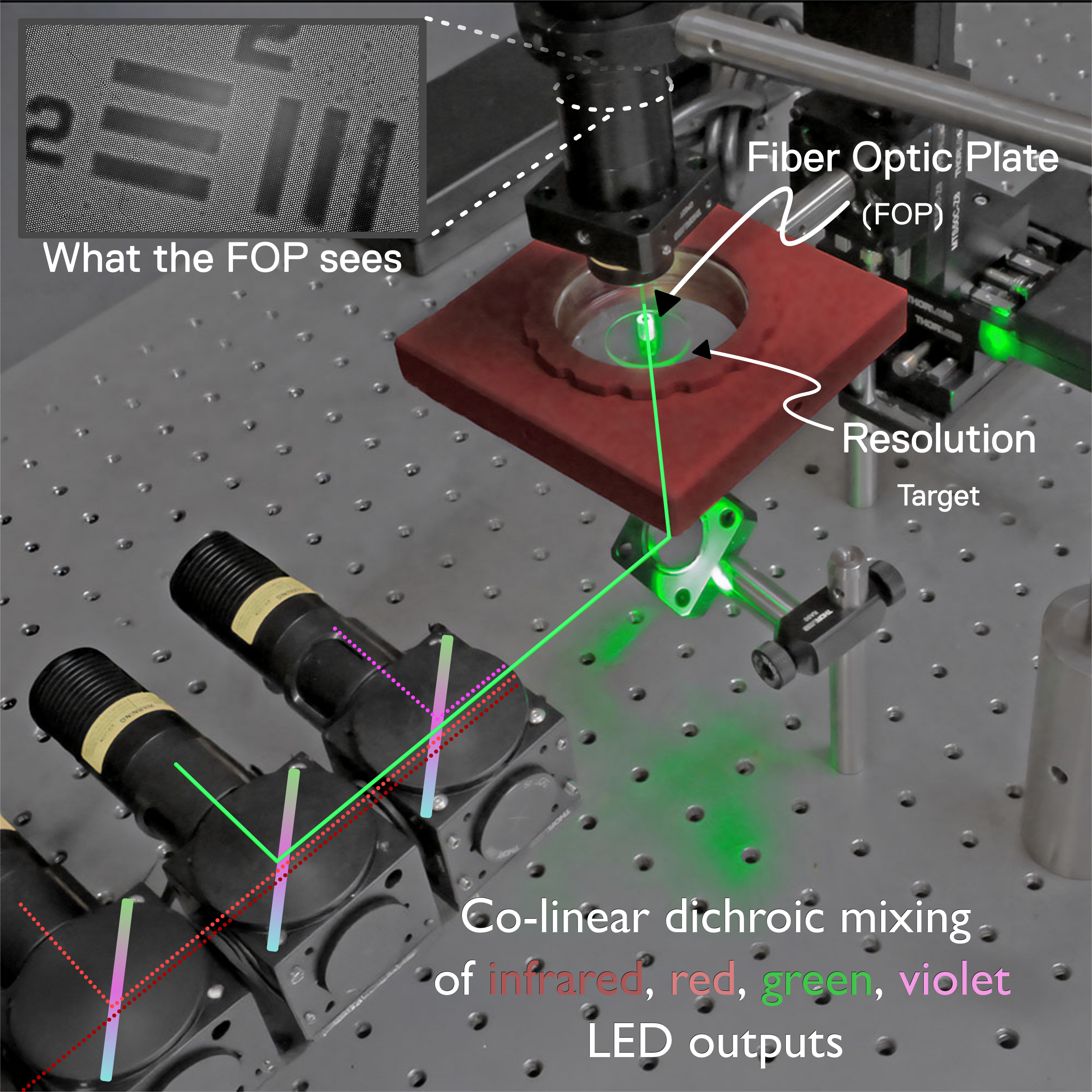
The image to the left depicts a laboratory setup that benchmarks the performance of our FOP prototypes, including mounts, lenses, scattering samples, and illumination sources, across visible and near-infrared bands relevant to biological imaging. We have introduced tissue-mimicking phantoms to simulate turbid media and depth conditions, enabling comparison of signal quality across increasingly challenging optical paths. The FOP is often placed on a known resolution target and evaluated quantitatively and visually to compare prototypes. The samples are assessed for resolution, contrast, spatial fidelity, edge sharpness, brightness uniformity, and interrogation depth, which together indicate how reliably the FOP preserves image information beneath scattering layers.
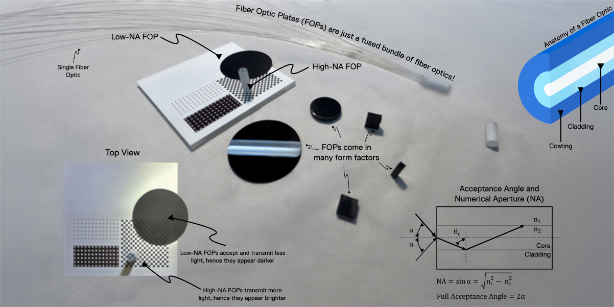
Fibre-optic plates (FOPs) are essentially fused bundles of thousands of tiny optical fibres, so they inherit the same basic light-guiding principles as ordinary fibre optics. Each fibre has a core, cladding, and coating, and its ability to accept and transmit incoming light is governed by properties such as numerical aperture (NA), which defines the range of angles over which light can be coupled into the fibre. A high-NA FOP accepts more light and therefore appears brighter, while a low-NA FOP accepts and transmits less light, making it look physically darker. However, this reduced acceptance angle can be useful for imaging through turbid media, as it rejects and suppresses highly scattered light and preferentially collects photons that remain mainly unscattered, emerging from deeper within the sample. In this way, lowering the NA can trade brightness for improved depth selectivity and potentially increase the effective interrogation depth.
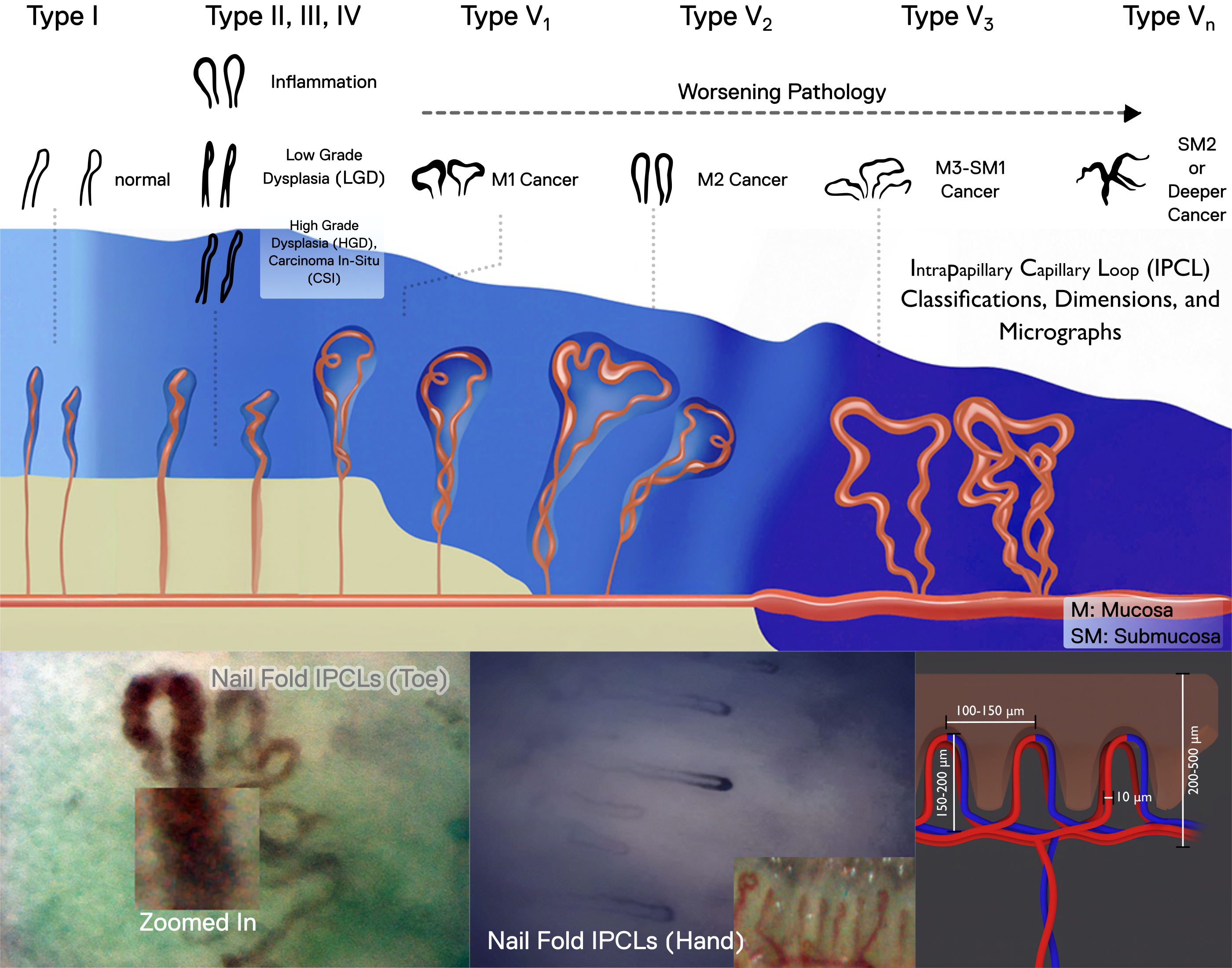
FOPs can serve as an optical interface for assessing deeper intrapapillary capillary loops (IPCLs), especially in turbid tissues. The spatial angular filtering enabled by FOPs can suppress shallow, multiply scattered photons and improve visibility to deeper structures. IPCL morphologies are clinically important; as tissue progresses from normal mucosa toward superficial cancer, IPCLs can become dilated, branched, tangled, irregular, and variable in shape. Therefore, FOPs can serve as validation tools for earlier detection and future risk prediction for superficial cancers.