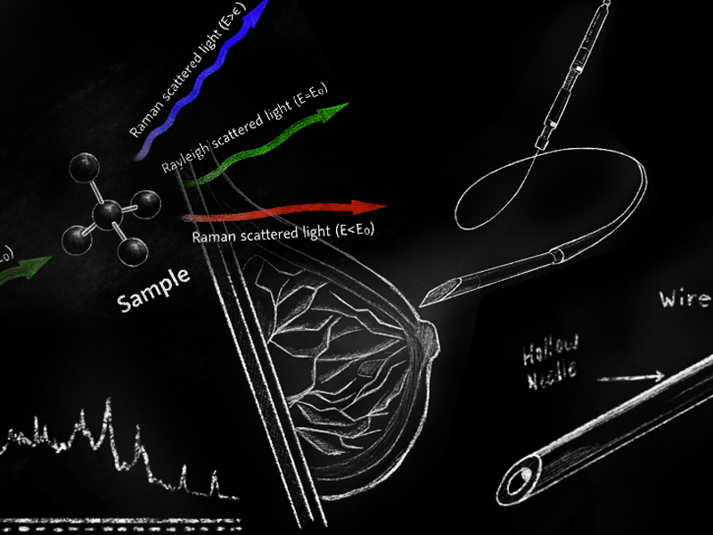
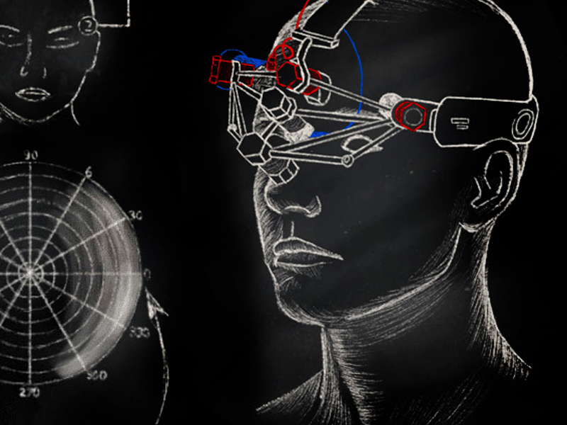
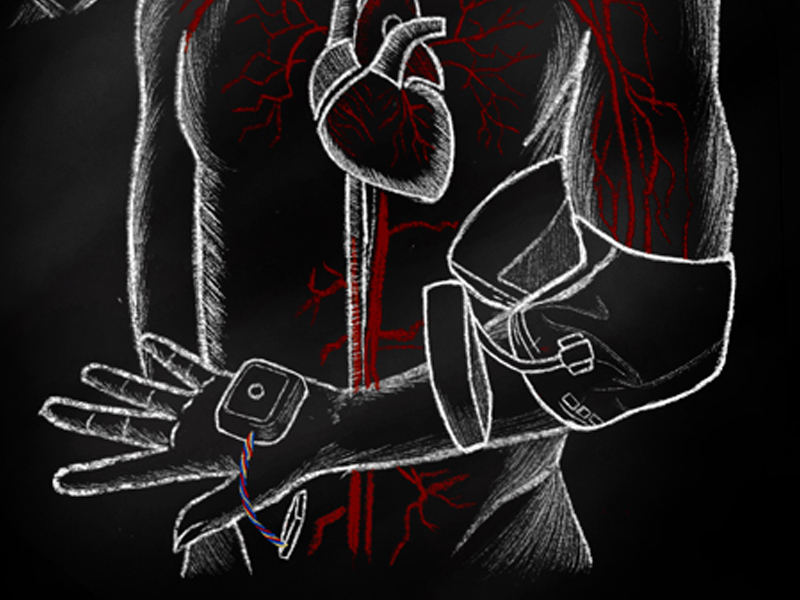
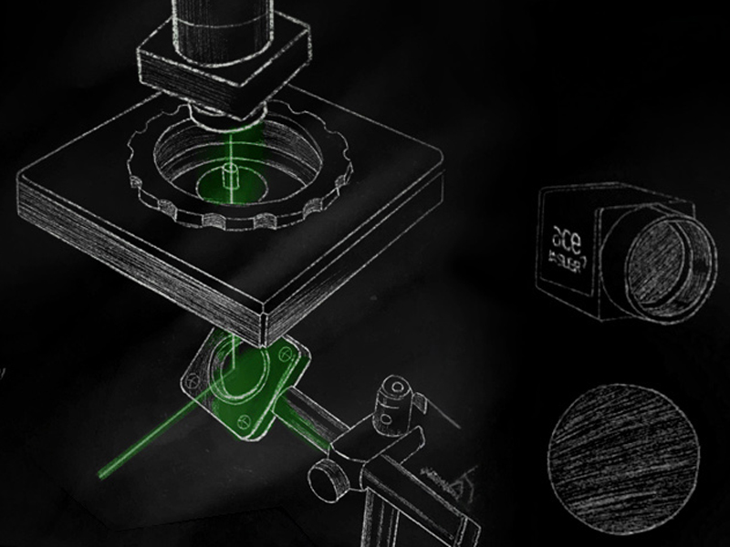
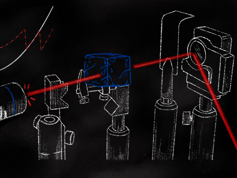
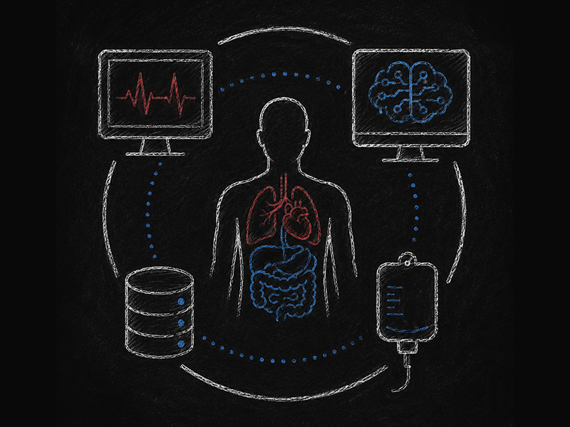
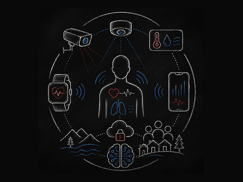
You are now in the main content area

Research Projects

The project is devoted to development of a microendoscope, which outer diameter is 195 micron, the inner diameter is 100 micron, and the length is 45 mm. This outer diameter is comparable with an acupuncture needle, but our needles are hollow and can deliver an endoscope. The main advantage of our scope - it can take small biopsies under imaging and spectroscopy control practically painless and causing no bleeding. The tissue destruction is minimal and healing is fast. It gives an opportunity to perform biopsies and collect diagnostic data not only in hospitals under anesthesia, but also in small clinics and to apply biopsy easier obtaining considerably more data per patient. This is the main point why such an endoscope is a game changer for the patient care, opening much more robust diagnostics prospective. An optical schematics and fluorescence imaging of normal and cancer in breast can be seen in the pictures. This is a collaboration project with University Health Network, Mount Sinai Hospital and Ontario Cancer Centre.

Interstitial Needle Microendoscopy (Color Image, Fluorescence, RAMAN): Mosquito Bite Needle Endoscopy (MBNE)

The project is devoted to development of a deep optical imaging technique overcoming the blurring effect due to the high light scattering in biological tissues.  A spatial angle filter (SAF) has been invented. The recent generation of SAF facilitates a significant increase of the sampling/interrogated volume by 2-3 folds in tissues, which highly scatters light, such as skin. As the SAF technology does not require any mechanical/scanning or extra computational facilities, it can provide stable imaging collection in real time (live imaging). To date, to the best of our knowledge, there are no technologies on the market that facilitate deep skin imaging scanning with such high resolution at so low cost. Douplik’s group currently works on developing a compact skin scanner probe, similar to ultrasound probe, that can facilitate a quick cancer screening. A prototype implementation and experimental results of increasing sampling/interrogated volume by 2-3 folds in tissue intralipid phantoms can be seen in the pictures.

Spatial Angle Filter (SAF) Imaging: Seeing Deeper Inside Highly Light Scattering Media: Fog or Skin

The project is devoted to development of applications for Holography Interference Imaging, which is also called a double exposure holographic interferometry. This method facilitates a 2D map of interferences of the recorded and live holograms, and thus provides a very high accuracy of the spatially resolved light phase shift measurements in real time.  As shown in the figures, the project involves advanced phase wrapping and unwrapping technique towards various applications such as photochemical/photodynamic monitoring or dosimetry in radiology. This project is developed in collaboration with Ukrainian State University, Kyiv, Ukraine and Montpellier University, France.

Holography Interference (HI) Imaging

Precision Phosphene Control through Cutaneous Facial Electrical Stimulation

The project is devoted to application of a weak spatially resolved pulsating electrical currents across regions of the facial skin causing visual percepts experienced by the recipient called phosphenes (visually perceived flashes that are not caused by actual light). This is a non-invasive wearable technology as shown in the pictures. Through the strategic electro-stimulation of the phosphenes, a person can precisely control their location and flashing frequency in the field of vision. This method can be utilized in a visual prosthesis to aid the partially blind or visually impaired individuals to navigate the world if the phosphene stimulated device will be connected to the camera and/or global positioning system. This approach also can be used in computer gaming for interactive deep immersion.

PRECISION PHOSPHENE CONTROL THROUGH CUTANEOUS FACIAL ELECTRICAL STIMULATION, Alexandre Douplik, Ryerson.

Remote Cuffless Blood Preassure (BP) Monitoring

The project is devoted to development of remote touchless monitoring of blood pressure, pulse wave propagation velocity, heart rate, heart rate variability and respiratory rates. This technology is important for ICUs, long care facilities, clinics during pandemic where breathing and circulatory conditions are critical to control. The hardware/software prototype that was created can perform monitoring 24/7, even overnight exploiting a low power near infrared light source. This project is progressing in collaboration with Kagoshima University Hospital and Hamamatsu Corporation, Japan. The example of data collection of spatially resolved 2D pulse wave monitoring is shown: Pulse Transit Time (PTT) is determined as the maximum of the cross correlation between 2 segments of tissue to calculate Pulse Wave propagation velocity (PWV) and the blood pressure is computed via further transforms of the data.


REMOTE CUFFLESS BLOOD PREASSURE BP-MONITORING, Alexandre Douplik, Ryerson.

Fiber Probes for Remote Surface Enhanced RAMAN Spectroscopy Sensing

The project is devoted to development of fiber probes for Surface Enhanced Raman Spectroscopy (SERS). A custom designed Raman Spectroscopy collection setup was created for optimal acquisition of spectra via the optical fibers by building the plasmonic structure on the fiber tip employing the nanotechnological approach. The periodic pattern on the fiber tip is shown in the left sided figures. As a result of this work, we have developed the probes that currently facilitates 1-2 orders of magnitude of the Raman signal enhancement. Some results are shown in the right plot. The project is collaboratively supported by Tornado Spectral System, Toronto.

FIBER PROBES FOR REMOTE SURFACE ENHANCED RAMAN SPECTROSCOPY SENSING, Alexandre Douplik, Ryerson.

Biooptics 3D Phantoms

The project is devoted to development of phantoms to replace live objects for the experimental work in biooptics and optoacoustics including calibration and instrument specific diagnostic bias mitigation. We developed technology to simulate optical and acoustic properties of biotissues, including dynamic blood microcirculation and even heart beating. A unique technology of human hemoglobin containing polymers has been developed. Examples of bio phantoms and phantom mice are shown. We study a photodynamic reaction on a hemoglobin containing phantom, and a clear footprint of the photodynamic hemoglobin deoxygenation reaction after laser irradiation can be found in the figures.

BIOOPTICS 3D PHANTOMS, Alexandre Douplik, Ryerson.

Non-Destructive Quality Assesment of Stored Red Blood Cell Concentrates (RCC) Using Optical Spectroscopy

The project is devoted to development of non-destructive quality assessment of stored red blood cell concentrates (RCC) that will allow a blood service to monitor red blood cell (RBC) quality during storage without breaching sterility of the bag. We apply optical methods based on measuring the absorption and scattering of light without breaching RCC bag sealing identifying damage to the red blood cell membrane and alteration of the cell metabolic state that lead to red blood cell (RBC) physiological function losses. Four critical parameters are monitored: SO2 – blood oxygenation, HS – hemolysis, HA – hemoglobin amount, MA - RBC cell membrane alteration (volume fraction of spheroechinocytes). RCCs pictures, hemoglobin form spectra and preliminary results are shown in the figures. This project is developed in collaboration with Canadian Blood Service and University of Alberta (Prof. J. Acker).

NON-DESTRUCTIVE 
 QUALITY ASSESSMENT OF STORED RED BLOOD CELL CONCENTRATES, Alexandre Douplik, Ryerson.

The project is devoted to development of compact non-invasive red blood cell (RBC) counter that would not require drawing blood and rely upon image collections and sophisticated image processing. We develop a unique calibration method that can facilitate the RBC counts per ml totally non-invasively.

Counting Blood Cells, Prof. Alexandre Douplik, Ryerson University

The project is devoted to development of UVC/blue light system for disinfection of aircraft passenger cabin in combination with air ionization. This project is developed in collaboration with Department of Aerospace Engineering, Ryerson University - Ryerson Aerospace Engineering Centre (RAEC) (Prof. J. Xi) and Bombardier, Canada.

Development of UVC/Blue Light Disinfection for Aircraft Cabin