Hemodynamic Sensing
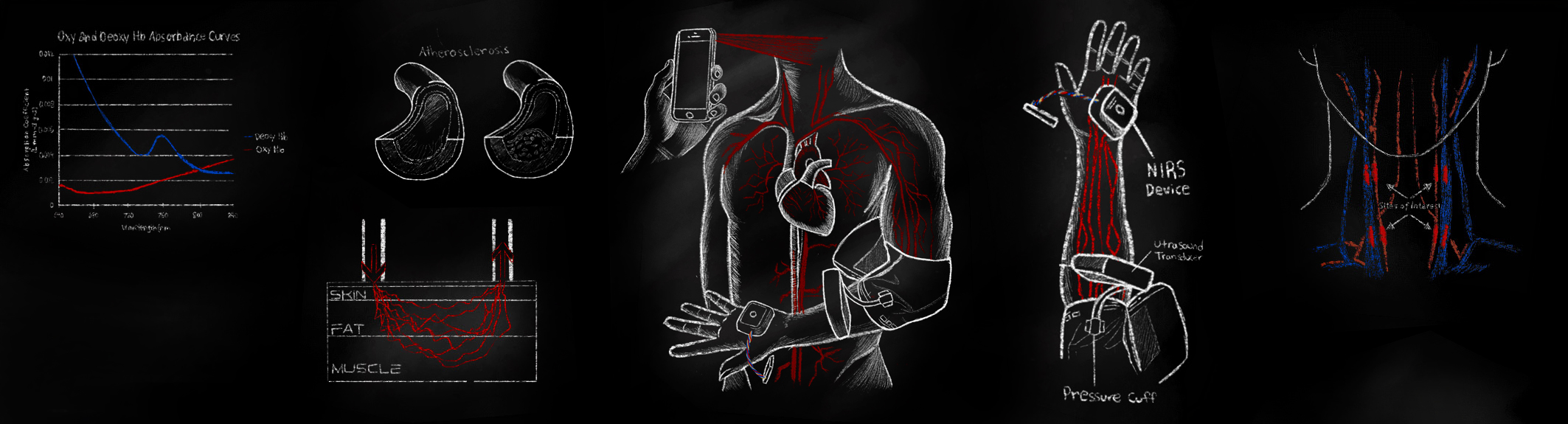
The project is dedicated to the development and exploration of both remote and contact instrumentation for measuring pulse wave velocity, local perfusion, heart rate, and muscle and blood oxygenation. This technology is crucial for ICUs, long-term care facilities, hospitals, and clinics, where circulatory conditions require precise monitoring.
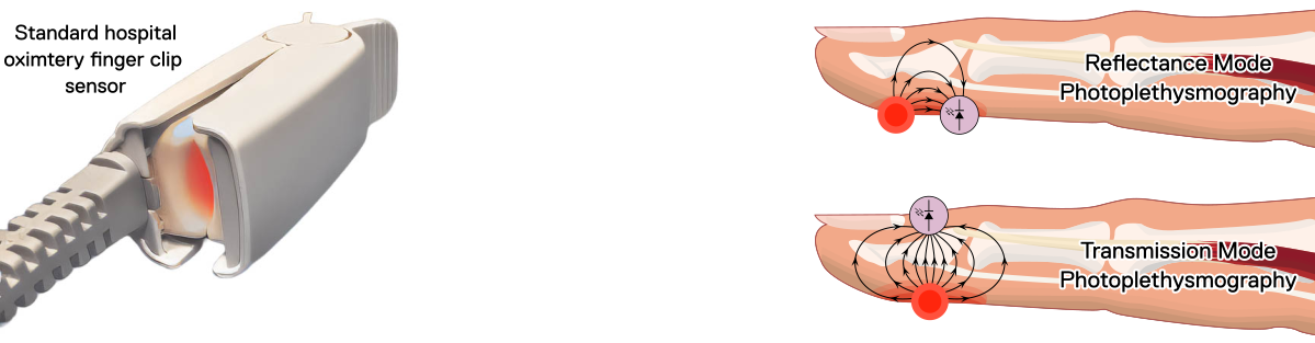
Our lab has a broad interest in this field, with many concurrent topics under investigation. Photoplethysmography (PPG) is a well-established technology first pioneered in the late 1930s. In general, PPG senses local changes in blood volume within tissues. Pulse oximetry, a PPG technology spin-off, uses multiple wavelengths of light to measure the blood oxygen saturation. This research led to the ubiquitous pulse oximeters found in hospitals and clinics, a critical health monitoring technology. The figure below depicts a common clinical fingertip pulse oximeter. Both PPG and pulse oximetry can be performed in transmission and reflectance modes. The mode depends on the placement of the main elements: the light emitter and the light sensor.
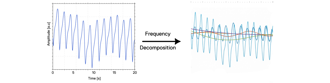
Photoplethysmography waveforms carry a rich mixture of physiological information. They not only convey changes in blood volume, but also multiple overlapping processes occurring at different time scales. The following diagram shows the physiological processes captured in a PPG waveform, decomposed and separated into their respective frequency bands.
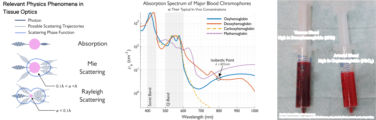
Understanding this field at a deeper level requires understanding the physics of how light interacts with biological tissue. The genesis of the PPG signal is primarily governed by absorption and scattering phenomena. Blood chromophores, most prominently oxyhemoglobin and deoxyhemoglobin, preferentially absorb specific wavelengths of light, governed by their absorption spectra. PPG and oximetry systems typically use red and infrared light because the differential absorption between these chromophores enables estimation of oxygen saturation (SpO₂). In short, this science models how raw light measurements translate into meaningful physiological insights. The following diagram depicts key photonic physical phenomena that define light-tissue interaction during PPG measurements. In the middle, the wavelength-dependent absorption spectra of the main blood chromophores are shown. On the right, a mind-blowing picture of arterial (oxyhemoglobin-rich) and venous (deoxyhemoglobin-rich) blood from the same person, with their striking colour differences, showing the stark effects of blood-chromophore concentration ratios on overall blood colour.
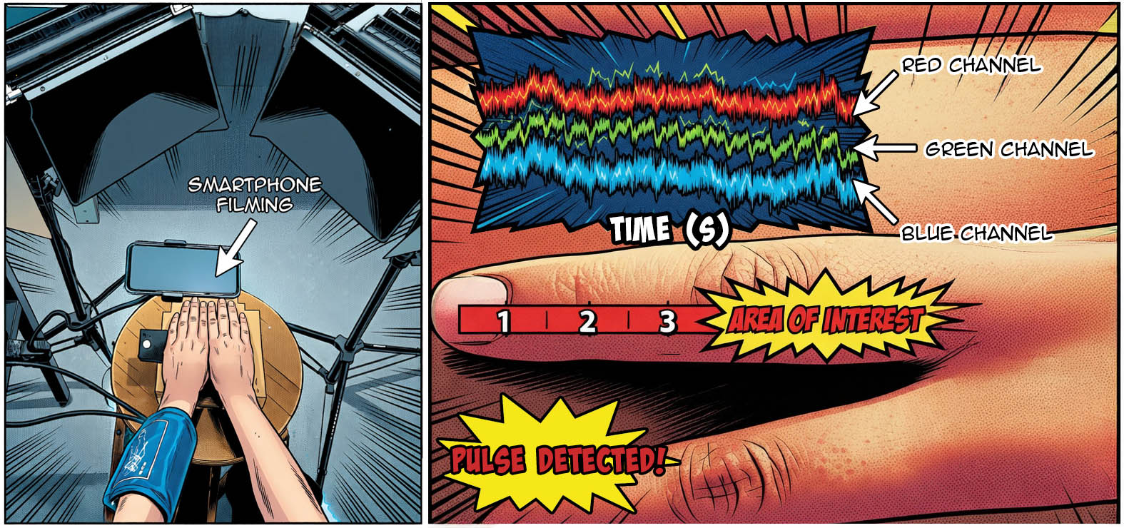
PPG and oximetric technologies mainly require a direct physical contact sensor to be applied to a human subject, which may significantly limit their overall versatility and broader practical applications. As a remedy, we also explored remote photoplethysmography (rPPG) as a non-contact alternative optical method for accurately detecting underlying cardiovascular dynamics. A simple consumer-grade smartphone camera can be effectively used to videograph regions of exposed skin to capture meaningful physiological signals. From subtle changes in skin colour over time, we can extract cardiac-cycle-associated pulsatile waveforms from the recorded video signal's colour channels. The green channel, particularly, consistently yields the best signal quality, due to hemoglobin’s distinct absorption spectra.
As seen in the figure above, in one study, we introduced an intervention: one arm was occluded with a pressure cuff, while the other served as a control. In the control arm, the rPPG signal exhibited clear cardiac-cycle pulsations. Conversely, the cuffed arm showed attenuation or an absence of the pulsatile waveform as the blood flow was restricted. This study demonstrated the value of rPPG in monitoring tissue perfusion, a critical health metric in clinical settings.
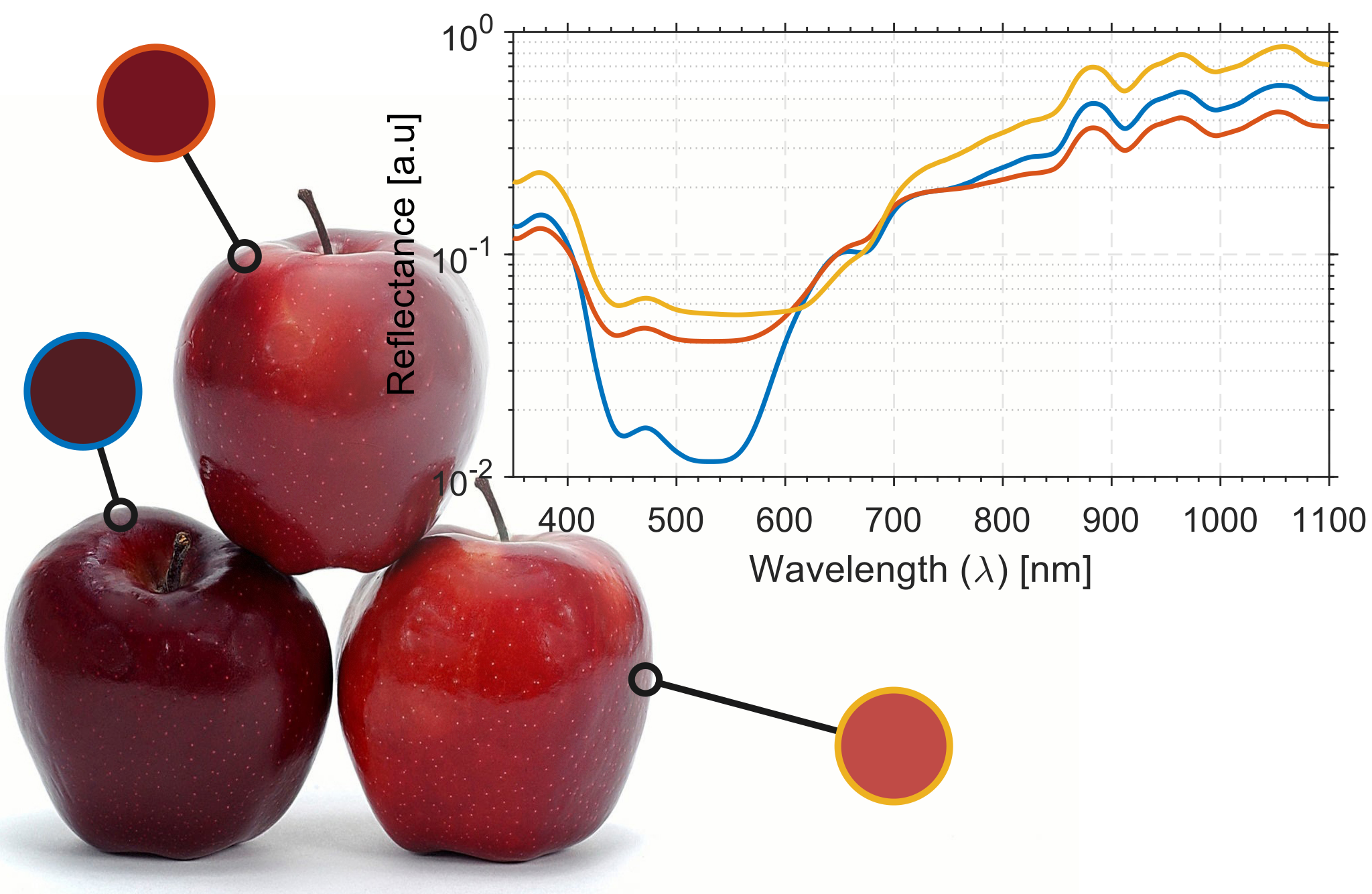
The study of absolute colour and the longer-term changes in skin colour has also sparked our interest. As shown in the figure below, apples of the same variety exhibit slight discrepancies in peel colour and reflectance spectra due to natural variations in ripening, resulting in small differences in peel chromophore concentration. Hence, colour analysis should be able to extract some information about chromophore concentrations in a biochemical environment. Similarly, the same principle and approach can be applied to quantitatively analyze a person's skin colour to extract key health metrics. Conditions such as physiological shock, anemia, mottled skin, hypoxia, and Raynaud's syndrome manifest symptoms that alter skin colour. Hence, this technology can help longitudinally monitor patients in hospitals and clinics with limited staffing.
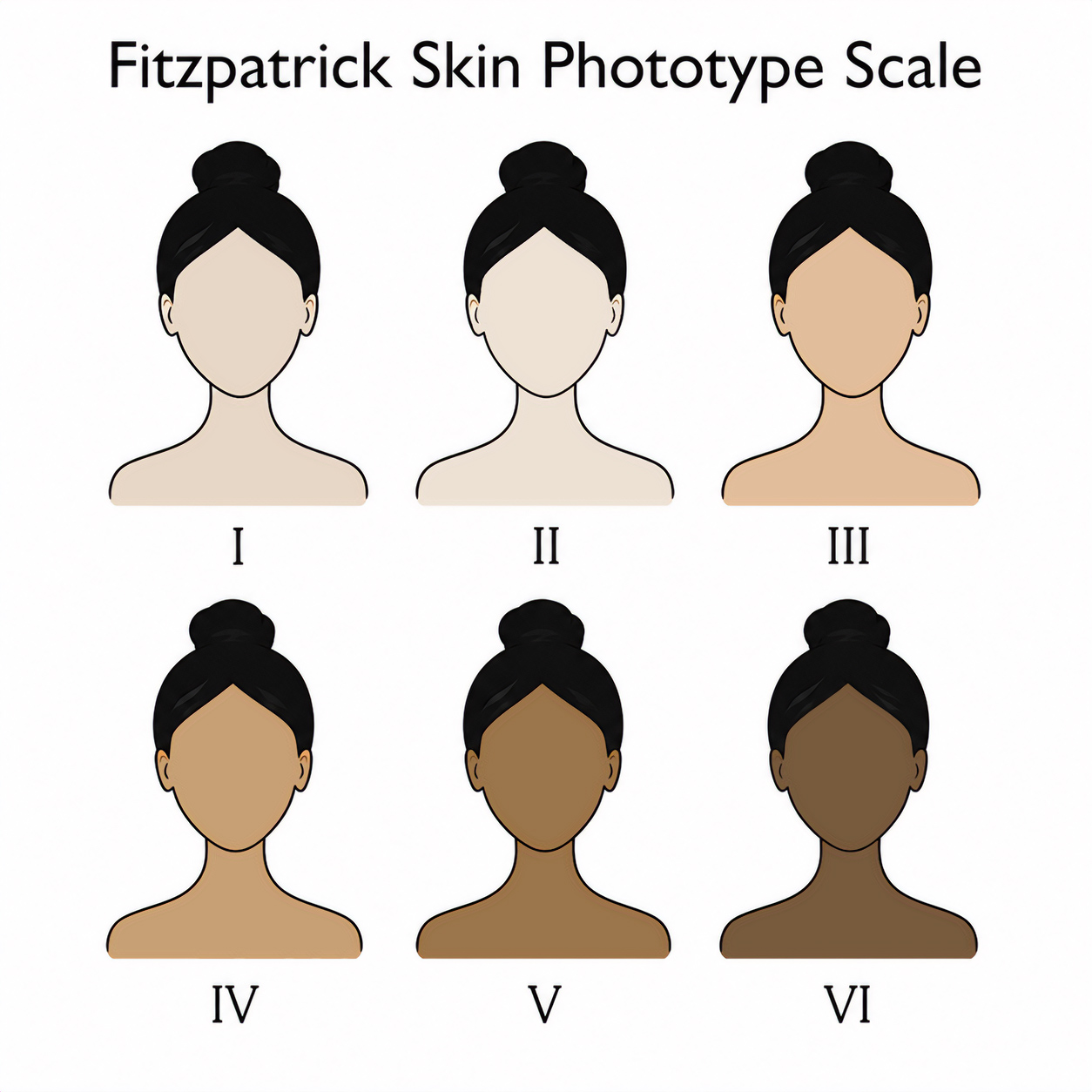
Furthermore, ambient lighting and differences in skin colour can greatly confound colour analysis in non-laboratory conditions. Hence, sophisticated algorithms are needed to adjust and calibrate colour sensing efforts to make accurate assessments of one's health metrics. The broad variation in baseline skin colour across the human population is by far the greatest technical challenge in biophotonic metrology.
This problem has inspired research and the development of algorithms that can universalize biophotonic measurements. The goal is to utilize computational simulations to model the effects of confounding factors on biophotonic signal characteristics. In our simulations, we use the Monte Carlo method, which excels at modelling probabilistic phenomena such as light transport. This method opens up the possibility of simulating a wide range of preconditions. The simulation output yields look-up tables that would significantly enhance measurement accuracy across a wide range of scenarios.
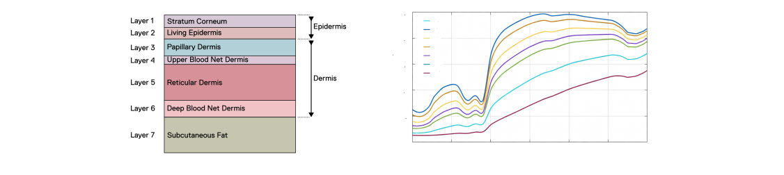
Monte Carlo methods improve both remote (rPPG) and contact photonic measurements by simulating how light scatters, absorbs, and propagates through layered tissues such as the epidermis and dermis. By modelling millions of photons and dialling in accurate tissue optical parameters, we can undo the effects of ambient lighting, skin tone, and geometry. This results in more accurate recovery of physiological signals, especially in complex or non-ideal measurement conditions. The following diagram shows an example of the Monte Carlo tissue-layering setup and the spectral effects of melanin concentration on light reflection from the skin.