Research
MFI Laboratory

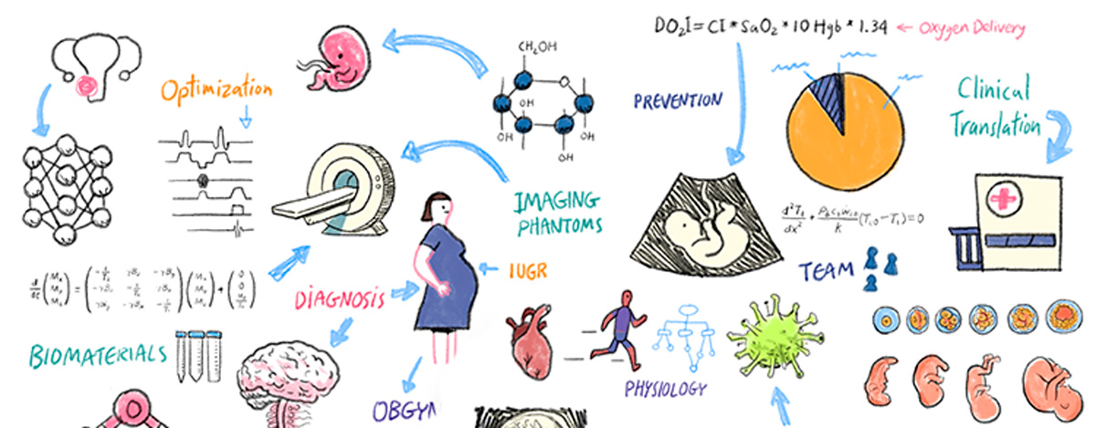
At the Maternal-Fetal Imaging (MFI) Laboratory, we develop and apply a range of biomedical engineering tools to facilitate the early diagnosis, treatment, and prognostication of obstetric and gynecological diseases. Our work spans the development of novel magnetic resonance imaging (MRI) sequences, machine learning models, fetal-placental imaging phantoms, and surgical technologies. We then translate these tools into clinical settings to improve our understanding of the physiology of health and disease in women, fetuses, and neonates.
Current Research
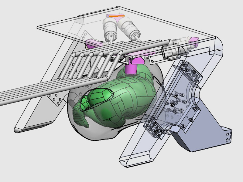
We are developing and testing robotic systems for use within the MRI machine to generate phantom motion during imaging, replicating artifacts that make gestational imaging challenging. These platforms enable researchers to optimize imaging sequences and develop more robust clinical scans.
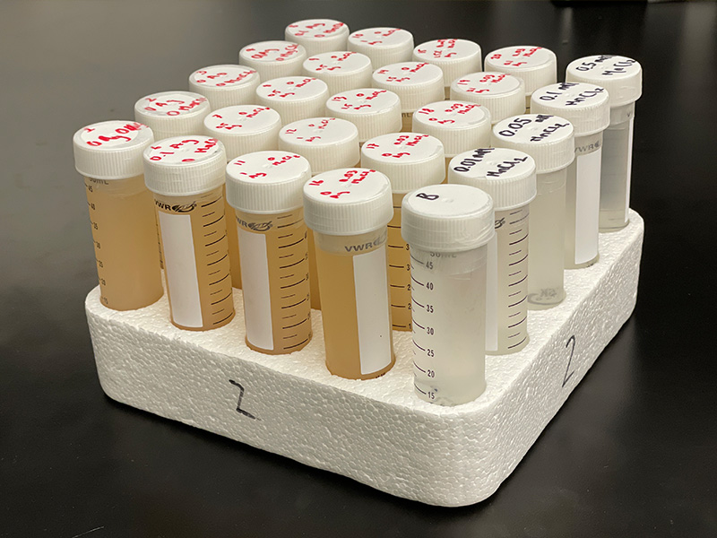
We are synthesizing novel materials that mimic the MRI properties of different human tissues.
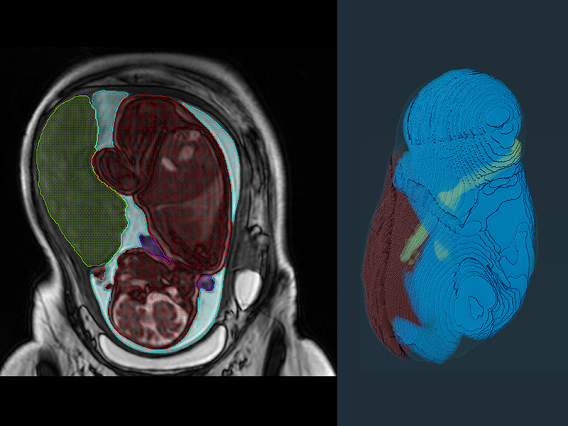
We are using machine learning approaches along with image processing techniques to develop novel algorithms for segmentation and disease classification of the placenta, fetus, and fetal organs from MR images. These will permit earlier and more accurate disease detection and facilitate interventions.
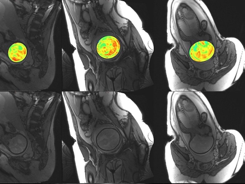
Our lab develops MRI techniques for non-invasive imaging of fetal and maternal physiology during pregnancy. These methods enhance our ability to investigate tissue metabolism, microstructure, and function, with applications in maternal screening and early detection of gestational complications. By employing approaches such as Saturation Transfer (ST) imaging, relaxometry, and Susceptibility-Weighted Imaging (SWI), our MRI techniques are sensitive to changes in tissue function that cannot be imaged with conventional clinical MRI. With these techniques, we aim to extend the capabilities of MRI in ways that support accessibility and clinical translation.
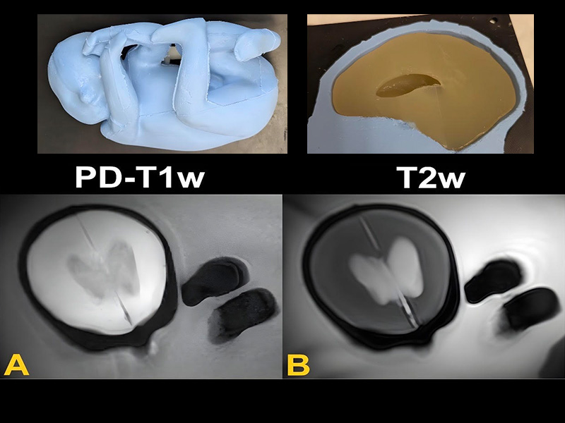
We are synthesizing 3D phantoms of the human fetus for magnetic resonance testing. This phantom will enable us to optimize various MR sequences as well as motion correction algorithms.
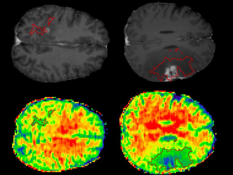
Clinical surveillance of brain tumours is typically a manual task aimed at assessing tumour growth and transformation. We are working closely with neurosurgeons and radiologists to develop an algorithm that automatically carries out tumour grading from non-contrast enhanced 3D MRIs.
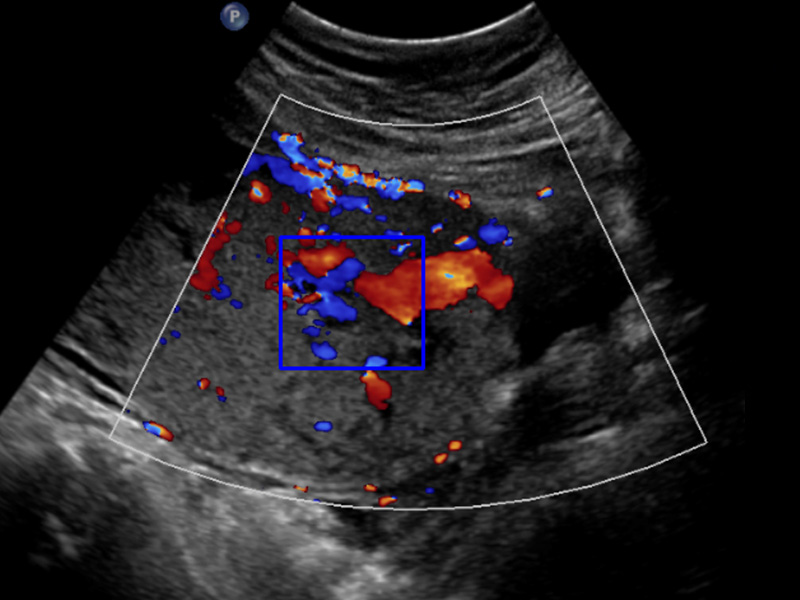
We are developing machine learning algorithms for detecting and classifying placenta accreta spectrum (PAS) disorder, where the placenta invades the myometrium. This will allow for earlier and more accurate diagnosis, facilitating clinical management and decreasing mortality.
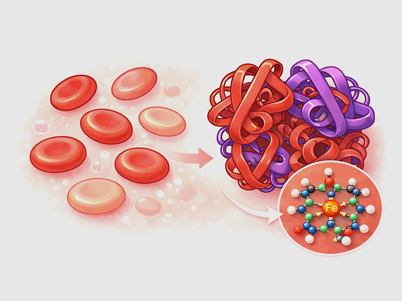
Many female patients are prone to iron deficiency (ID) secondary to heavy menstrual blood loss. While blood loss generally correlates with iron loss, individual perception and physiological factors complicate this relationship, making it challenging to predict ID. We are developing a non-invasive product to quantify iron loss during menstruation.
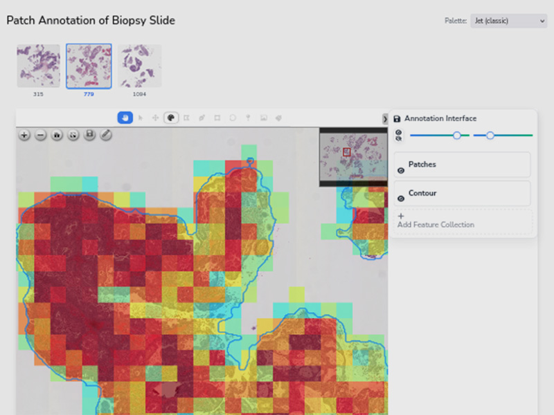
Endometrial biopsy is done in any female with abnormal uterine bleeding. While the majority of biopsies are benign, the large volume of cases delays diagnosis of endometrial cancer, which has a high survival rate when detected early. We are developing AI-based models for triaging of biopsy cases, rapid cancer detection and subclassification from pathology whole slide images. These tools are designed to improve workflow, expedite diagnosis, and facilitate earlier intervention.

Endometriosis presents with heterogeneous and non-specific symptoms, contributing to delays in diagnosis. We are developing patient-facing and clinician-facing AI-based models to improve patient referral pathways, early detection and disease staging altogether aiming to improve endometriosis healthcare.
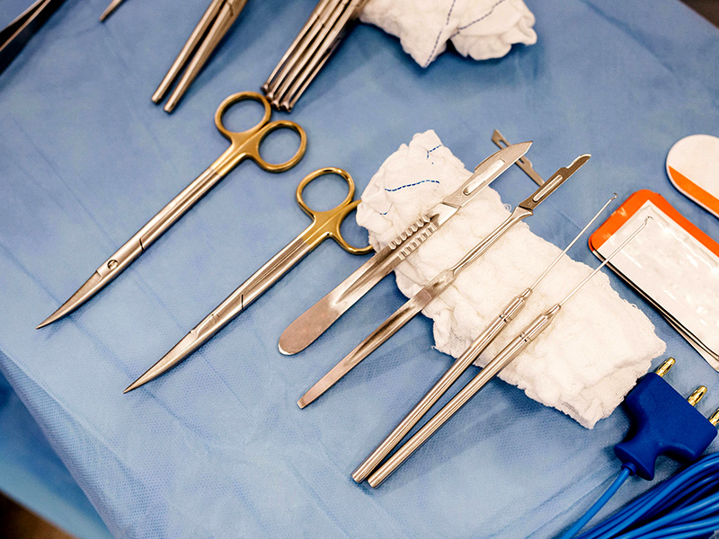
We are developing new techniques for medical learners to develop and practice their surgical skills and receive constructive feedback from their educators.