TMU students build AI tool to detect brain tumours
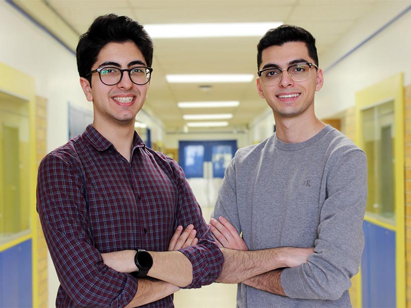
TMU students Kamyar Saeedabadi (left) and Daniel Dabir (right) are working on a brain tumour detection project that uses machine learning to provide more accurate and efficient brain tumour diagnoses.
Daniel Dabir and Kamyar Saeedabadi have been friends since a young age and share a passion for science and technology. With the rapid development of artificial intelligence (AI) in the last few years, they became interested in exploring how machine learning could improve the health care system.
“AI is becoming increasingly accessible nowadays and we see it being implemented in almost every aspect of our lives,” said Saeedabadi, who is studying industrial engineering. “So we thought, why not implement something that can help make a huge impact with our current health care system?”
The two TMU students are now spearheading a project that uses machine learning to detect and classify brain tumours. Their tool aims to meet three primary goals: decreasing wait times for radiologists, reducing health care costs and improving medical outcomes in hospitals and health care settings.
“The Canadian Association of Radiologists (PDF file) published data (external link) showing that in 2022, the average person would wait 89 days for an MRI and this wait time cost the industry approximately five billion dollars,” psychology student Dabir said. “Our idea was to increase efficiency and reduce wait times, which would result in increased cost savings. By using this AI-powered tool, we can improve the speed and accuracy of MRI scans, and see many beneficial outcomes.”
April Khademi, Canada Research Chair in AI for Medical Imaging, and biomedical engineering professor at TMU, says AI tools for medical imaging hold promise in improving diagnostic accuracy and turnaround times for patients suffering from neurological disease.
“Using AI for brain tumour diagnosis and management could lead to more personalized therapeutic regimes which ultimately improve quality of care. Additionally, AI tools can lead to faster diagnostic turnaround times which reduces stress on the patients,” Khademi said.
Easy-to-use technology for increased adoption
The tool enables health-care professionals to upload an image or MRI scan – and conducts an image analysis to accurately classify the tumour in one of four categories: meningioma, glioma, pituitary and non-tumour (i.e., a clean MRI scan).
In designing their tool, Dabir and Saeedabadi used a deep learning algorithm called Convolutional Neural Network, that analyzes the images you input and processes them using mathematical operations.
“The Convolutional Neural Network is widely available and people use it for various things – it has many implications in health care that people are now starting to tap into,” Dabir said.
“The Canadian Radiology Association’s report recommended that an AI or deep learning machine should be built to aid radiologists. The possibilities are endless because it's mostly just the classification of images – and that’s really the entire principle of radiology – whether it’s scans of lungs or broken bones or bleeds in the body. But we put our own twist on it by using it for MRI scans.”
Khademi added that AI will revolutionize the way physicians practise by providing them with new, quantitative metrics of disease for a variety of modalities. “While we are in this rapid technology evolution phase, it is important for physicians to be involved in the design as the ultimate end-users,” she said.
Work in progress
To make the tool as accessible and easy to use as possible, the students adapted the model to Google’s Teachable Machine (external link) so that anyone can input an image from their own scans and get a result. However, with greater ease come a few limitations.
“You need a great data set to provide fully accurate results, and there are variations in tumour location, shape and size, which can skew results and diagnostics,” said Saeedabadi. “But the technology is still a great tool that can aid the diagnostic process and flag certain MRIs. As with any technology, there's always room for improvement when it comes to design and testing.”
The students are currently working on broadening the dataset, prioritizing increased accuracy to enhance the tool. They are also in the process of drafting proposals for potential research partnerships with health care organizations.
“We are optimistic about the potential partnerships and the impact our project could have on the field of medical diagnostics. We are initiating discussions with medical institutions in the Toronto area to explore potential collaborations and opportunities for implementing our AI-based system in real-world medical settings,” Dabir said.